Celastrol (Tripterine) Attenuates Ischemia-Reperfusion Injury by Suppressing the HIF-1α/PDK1 Axis
Copy code to add this image to your page
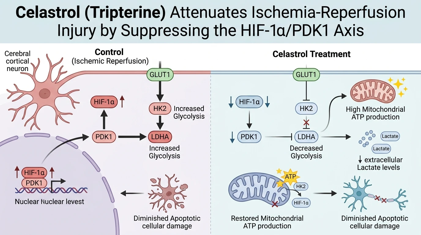
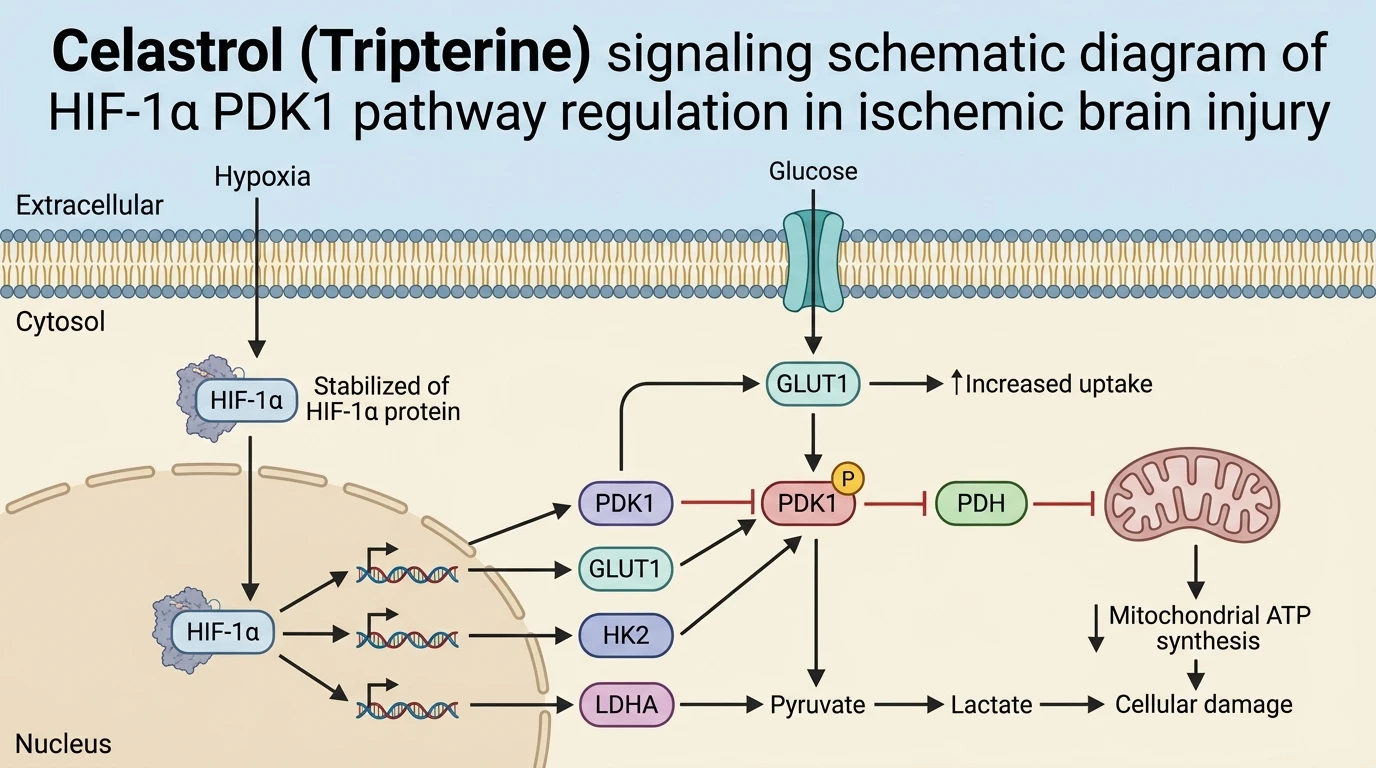
Cerebral ischemia/reperfusion (I/R) injury triggers a profound disruption in cellular energy homeostasis, forcing neural tissue into acute anaerobic glycolysis. While emergency glycolytic engagement initially maintains minimal ATP production during hypoxic stress, sustained glycolytic activation leads to intracellular toxic acidosis and metabolic collapse. Central to this hypoxic metabolic adaptation is the hypoxia-inducible factor 1-alpha (HIF-1α) and pyruvate dehydrogenase kinase 1 (PDK1) signaling axis. Upon ischemic onset, stabilization of HIF-1α upregulates PDK1 expression, which phosphorylates pyruvate dehydrogenase (PDH) to prevent pyruvate entry into the tricarboxylic acid cycle. Consequently, cells exhibit elevated expression of downstream glycolytic machinery—including glucose transporter 1 (GLUT1), hexokinase 2 (HK2), and lactate dehydrogenase A (LDHA)—causing abnormal lactate accumulation and mitochondrial breakdown.
Evaluation of natural pentacyclic triterpenoids revealed that Celastrol (Tripterine) acts as a potent small-molecule modulator of metabolic flux during ischemic stress. Investigated within a murine model of transient middle cerebral artery occlusion (tMCAO), administration of Celastrol (Tripterine) yielded robust neuroprotective effects against focal brain ischemia. Systemic treatment reduced cerebral infarct volumes in a clear dose-dependent manner. Cognitive and motor testing showed marked improvements in neurological deficit scores. Dose-optimization assays identified 4.5 mg/kg as the optimal therapeutic window in mice, balancing efficacy with safety. Histological analysis of ischemic cortical tissue showed a notable reduction in TUNEL-positive apoptotic neurons, confirming that the structural integrity of the cortical mantle was preserved under treatment.
Mechanistic dissecting of tissue lysates demonstrated that Celastrol (Tripterine) targets the transcriptional control of cerebral bioenergetics. Treatment blocked the hypoxia-induced upregulation of HIF-1α protein levels and suppressed downstream PDK1 expression in the ischemic cortex. This suppression effectively uncoupled the pathological drive toward excessive glycolysis. Western blot analysis confirmed significant reductions in key rate-limiting enzymes: GLUT1 expression fell, preventing unregulated glucose uptake; HK2 expression decreased, normalizing initial hexose phosphorylation; and LDHA abundance declined sharply. Concurrently, lactate concentration within ischemic tissue decreased, relieving localized tissue acidosis while cellular ATP levels and free glucose concentrations were restored toward baseline physiology.
To establish direct target engagement, researchers introduced the prolyl hydroxylase inhibitor dimethyloxalylglycine (DMOG), a pharmacological activator of HIF-1α. Co-administration of DMOG effectively stabilized HIF-1α even in the presence of the active compound. DMOG co-treatment completely reversed the suppression of PDK1, GLUT1, HK2, and LDHA, driving lactate accumulation back to pathological levels. Crucially, HIF-1α activation abated the compound's neuroprotective benefits, causing a rebound in infarct volume and functional neurological impairment. These rescue experiments establish that Celastrol (Tripterine) confers neuroprotection primarily by disabling the HIF-1α/PDK1-mediated glycolytic cascade.
Pharmacological Characterization and Metabolic Pathway Modulation
Copy code to add this image to your page
Celastrol (Tripterine) protects against cerebral ischemia/reperfusion injury by inhibiting the HIF-1α/PDK1 signaling axis. Downregulation of HIF-1α and PDK1 suppresses downstream glycolytic enzymes (GLUT1, HK2, and LDHA), lowering toxic lactate accumulation while restoring ATP and glucose levels. In mice subjected to transient middle cerebral artery occlusion, an optimal dose of 4.5 mg/kg reduced cerebral infarct volume and improved neurological function. Target engagement was verified when the HIF-1α activator DMOG completely reversed these neuroprotective and metabolic effects.
Attenuation of Ischemic Cortical Infarction and Neurological Deficits
In vivo characterization using a transient middle cerebral artery occlusion mouse model demonstrated clear dose-dependent therapeutic efficacy. Neurological deficit scores improved significantly within 24 hours post-reperfusion. Infarct volume decreased substantially following systemic treatment. Dose-response matrix evaluations established 4.5 mg/kg as the optimal therapeutic dose in murine subjects. At this concentration, Tripterine suppressed cortical neuronal death and preserved penumbral cytoarchitecture. Immunofluorescent staining revealed marked reductions in apoptotic markers across the ischemic core edge.
Suppression of HIF-1α/PDK1 Cascade Restores Cellular Bioenergetics
Ischemic conditions typically trigger severe metabolic dysregulation through stabilization of the HIF-1α transcription factor. Treatment with Tripterine effectively blunted HIF-1α protein accumulation in cortical tissue. Downstream expression of PDK1 dropped accordingly. This molecular inhibition disrupted the pathologically elevated expression of key glycolytic enzymes—specifically GLUT1, HK2, and LDHA. Parenchymal lactate levels decreased rapidly following enzyme suppression; cellular ATP pools recovered alongside normal glucose utilization within ischemic penumbral zones.
DMOG Activation Confirms Target Specificity of Glycolytic Inhibition
Target validation required direct pharmacological challenge using dimethyloxalylglycine, a classical HIF-1α hydroxylase inhibitor. DMOG administration successfully stabilized HIF-1α despite compound treatment. This intervention restored PDK1 expression and up-regulated downstream glycolytic enzymes. Lactate accumulation resumed in cortical tissue. Furthermore, DMOG reversed the reduction in cerebral infarct volume and abolished motor recovery. These rescue findings confirm that Tripterine targets the HIF-1α/PDK1 signaling axis to attenuate ischemic pathology.