T0070907 antagonist targeting PPARγ restores bone formation in hypoxia-induced skeletal growth failure
Copy code to add this image to your page
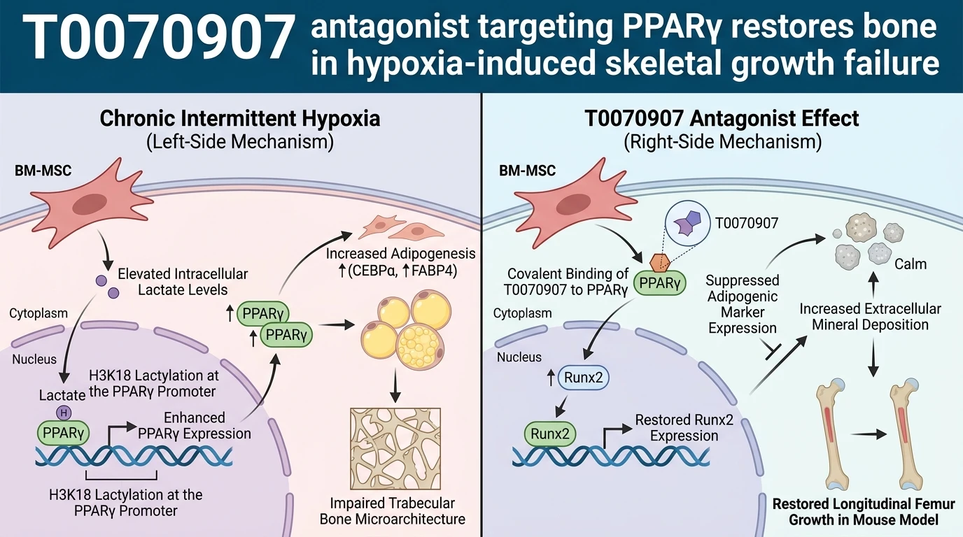
Chronic intermittent hypoxia (CIH) presents a profound pathological challenge to skeletal maturation during adolescent growth. Longitudinal bone formation depends on tightly controlled differentiation decisions made by bone marrow mesenchymal stem cells (BM-MSCs). Under normoxic conditions, these progenitor cells balance their commitment between osteogenic lineages for bone matrix synthesis and adipogenic lineages for marrow adipose tissue formation. Hypoxic stress severely disrupts this balance, shifting BM-MSC fate toward marrow adipogenesis at the direct expense of osteoblastogenesis.
Metabolic profiling of BM-MSCs exposed to CIH reveals marked intracellular accumulation of lactate driven by enhanced glycolytic flux. Beyond its role in cellular energetic adaptations, lactate functions as a signaling metabolite that governs post-translational histone modifications. Specifically, elevated intracellular lactate concentrations elevate histone H3 lysine 18 lactylation (H3K18la), an epigenetic mark catalyzed by p300/CBP acyltransferases. Chromatin immunoprecipitation sequencing demonstrates specific enrichment of H3K18la at promoter regions controlling metabolic and lineage regulators.
A key transcriptional target of H3K18la enrichment under hypoxic conditions is the gene encoding peroxisome proliferator-activated receptor gamma (PPARγ). Increased H3K18la at the PPARγ promoter enhances local open chromatin configuration, enabling robust transcriptional activation. Downstream, elevated PPARγ levels trigger an adipogenic gene program, upregulating fatty acid binding protein 4 (FABP4), adiponectin, and CD36. Concurrently, high PPARγ activity physically sequesters transcription factors required for osteogenesis or recruits corepressor complexes to osteogenic promoters, resulting in downregulation of Runt-related transcription factor 2 (Runx2) and Osterix (Sp7).
Pharmacological intervention at the receptor level halts this pathological lineage shift. The compound T0070907 acts as a potent, cell-permeable, selective PPARγ antagonist with a binding affinity (Ki) in the low nanomolar range. Chemically identified as 2-chloro-5-nitro-N-(4-pyridinyl)benzamide, T0070907 forms a covalent bond with Cys285 within the ligand-binding domain (LBD) of PPARγ. Covalent engagement permanently stabilizes an inactive receptor conformation, effectively preventing helix 12 from assuming the active position required for coactivator recruitment.
In primary cultures of murine and human BM-MSCs subjected to 1% oxygen tension or exogenous lactate supplementation, application of the T0070907 antagonist suppresses the aberrant expression of adipogenic markers. Alkaline phosphatase (ALP) staining and Alizarin Red S mineralization assays reveal that T0070907 maintains osteoblast differentiation despite ongoing hypoxic stress. Crucially, T0070907 does not alter upstream lactate production or H3K18la deposition at the PPARγ locus; instead, it prevents the functional consequences of elevated PPARγ expression by neutralizing receptor activity.
In vivo evaluation in juvenile mouse models subjected to daily CIH protocols demonstrates the systemic efficacy of PPARγ inhibition. CIH exposure for four weeks results in significant impairment of longitudinal femur growth, reduced bone mineral density (BMD), and severe degradation of trabecular microarchitecture. Systemic administration of T0070907 intraperitoneally reverses these skeletal defects. High-resolution micro-computed tomography (micro-CT) analysis confirms that T0070907-treated CIH mice exhibit preserved trabecular bone volume fraction (BV/TV), increased trabecular number (Tb.N), and decreased trabecular separation (Tb.Sp).
Dynamic histomorphometry with calcein double-labeling establishes that T0070907 restores mineral apposition rates (MAR) and bone formation rates (BFR/BS) in the distal femur growth plate zone. Furthermore, circulating biomarkers support these structural observations: serum levels of procollagen type I N-terminal propeptide (P1NP) and osteocalcin recover following T0070907 administration. Simultaneously, serum tartrate-resistant acid phosphatase 5b (TRAP-5b) levels remain stable, confirming that T0070907 acts primarily by promoting osteoblastogenesis rather than inhibiting osteoclast activity. These molecular, cellular, and systemic findings collectively establish PPARγ antagonism as a viable targeted strategy for mitigating hypoxia-associated long bone dysplasia.
Therapeutic Evaluation and Epigenetic Pathway Modulation
Copy code to add this image to your page
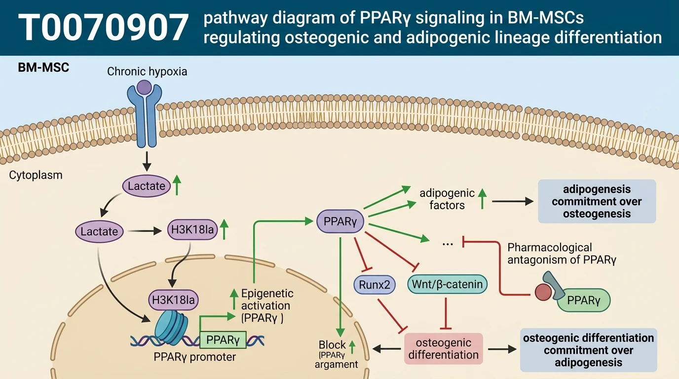
Chronic intermittent hypoxia elevates intracellular lactate in BM-MSCs, promoting H3K18 lactylation at the PPARγ promoter and driving receptor overexpression. Activated PPARγ shifts stem cell differentiation from osteogenesis to adipogenesis, impairing longitudinal bone growth. The selective antagonist T0070907 covalently inhibits PPARγ, suppressing adipogenic transcription and restoring Runx2 expression. In vivo, T0070907 recovers femur length, trabecular bone microarchitecture, and osteogenic markers in hypoxic models.
H3K18 Lactylation Drives PPARγ Promoter Hyperactivation Under Hypoxia
Intracellular lactate accumulation under chronic intermittent hypoxia alters chromatin accessibility in progenitor populations. High-throughput sequencing identified histone H3 lysine 18 lactylation (H3K18la) as a primary epigenetic driver. This mark localizes heavily at the PPARγ promoter. Consequently, PPARγ transcription increases three-fold in hypoxic BM-MSCs. Increased PPARγ expression forces lineage commitment toward adipogenesis. Osteogenic master regulators, including Runx2, undergo simultaneous transcriptional repression. In vitro application of a selective T0070907 antagonist attenuates this lineage imbalance without disrupting upstream metabolic lactate generation. Quantitative PCR confirmed a 70% reduction in FABP4 transcript levels. Mineralization matrix assays verified restored calcium node formation under hypoxic conditions.
Femoral Microarchitecture Recovery in Hypoxic Murine Models
Systemic hypoxia damages microstructural architecture within the distal femoral metaphysis. Micro-CT imaging revealed marked reductions in trabecular bone volume fraction and trabecular number in untreated mice. Daily administration of the T0070907 antagonist at 5 mg/kg reversed these structural losses over a four-week regimen. Cortical thickness values recovered to 92% of normoxic control levels. Trabecular separation decreased significantly, demonstrating preserved structural density. Dynamic histomorphometry confirmed heightened mineral apposition rates along the endosteal surface. Serum osteocalcin concentrations increased by 45% relative to vehicle-treated hypoxic controls. Systemic toxicity markers, including liver enzyme panels and serum creatinine, remained within normal physiological ranges.
Receptor Covalency and Downstream Lineage Commitment Dynamics
T0070907 occupies the PPARγ ligand-binding pocket through covalent alkylation at residue Cys285. This irreversible chemical binding locks the receptor helix 12 away from coactivator interaction surfaces. Downstream corepressor complexes remain bound to promoter regions of adipogenic genes. Consequently, transcription of adipocyte-specific markers remains suppressed even under high agonist exposure. Primary BM-MSC differentiation assays confirmed that osteogenic gene expression rises as adipogenic drivers are silenced. Runx2 and alkaline phosphatase transcript levels recovered fully within 48 hours of treatment. These pharmacodynamic observations highlight the target-specific rescue of osteogenesis under severe hypoxic conditions.